Figure 2
MALDI-MSI method for the detection of large biomolecules in plant leaf tissue
Lilian ST Carmo, Daiane G Ribeiro, Eder A Barbosa, Luciano P Silva and Angela Mehta*
Published: 06 August, 2021 | Volume 5 - Issue 2 | Pages: 058-061
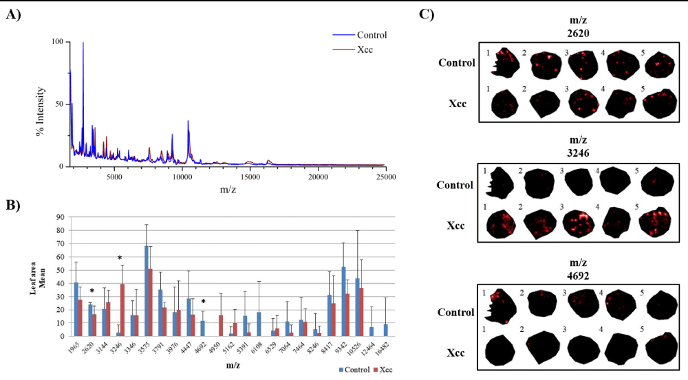
Figure 2:
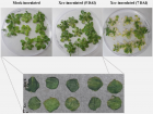
Potential protein biomarkers revealed in leaves of A. thaliana infected with Xanthomonas campestris pv. campestris (Xcc) using MALDIMSI approach. A) Overlapping image of the global mass spectrum obtained from the acquisitions of MALDI-MSI in the control and Xcc inoculated leaves. B) Graph of the leaf area mean for each ion. (*) Statistically validated ions by Student’s t-test (p-value ≤ 0.05). C) Ions distributed in leaf discs inoculated with Xcc and in the control condition. The numbers 1, 2, 3, 4 and 5 represent the biological replicates.
Read Full Article HTML DOI: 10.29328/journal.jpsp.1001061 Cite this Article Read Full Article PDF
More Images
Similar Articles
-
Role of polyamine metabolism in plant pathogen interactionsMagda Pal*,Tibor Janda. Role of polyamine metabolism in plant pathogen interactions. . 2017 doi: 10.29328/journal.jpsp.1001012; 1: 095-100
-
Role of HECT ubiquitin protein ligases in Arabidopsis thalianaYing Miao*,Wei Lan,Weibo Ma. Role of HECT ubiquitin protein ligases in Arabidopsis thaliana. . 2018 doi: 10.29328/journal.jpsp.1001016; 2: 020-030
-
Evaluation of cold response in Ilex paraguariensisCristian Antonio Rojas*,Sandonaid Andrei Geisler,Carina Francisca Argüelles. Evaluation of cold response in Ilex paraguariensis. . 2019 doi: 10.29328/journal.jpsp.1001026; 3: 009-012
-
Cloning and Characterization of a Pseudo-Response Regulator 7 (PRR7) Gene from Medicago Sativa Involved In Regulating the Circadian ClockWeifeng Nian*,Yilin Shen. Cloning and Characterization of a Pseudo-Response Regulator 7 (PRR7) Gene from Medicago Sativa Involved In Regulating the Circadian Clock. . 2019 doi: 10.29328/journal.jpsp.1001032; 3: 056-061
-
MALDI-MSI method for the detection of large biomolecules in plant leaf tissueLilian ST Carmo,Daiane G Ribeiro,Eder A Barbosa,Luciano P Silva,Angela Mehta*. MALDI-MSI method for the detection of large biomolecules in plant leaf tissue. . 2021 doi: 10.29328/journal.jpsp.1001061; 5: 058-061
-
Flashes of UV-C light are perceived by UVR8, the photoreceptor of UV-B lightJawad Aarrouf*,Douae Ben Hdech,Alice Diot,Isabelle Bornard,Lauri Félicie,Laurent Urban*. Flashes of UV-C light are perceived by UVR8, the photoreceptor of UV-B light. . 2022 doi: 10.29328/journal.jpsp.1001089; 6: 151-153
-
Snapshot of the Involvement of Glutathione in Plant-Pathogen InteractionsAparupa Bose Mazumdar Ghosh, Sharmila Chattopadhyay*. Snapshot of the Involvement of Glutathione in Plant-Pathogen Interactions. . 2023 doi: 10.29328/journal.jpsp.1001103; 7: 039-041
Recently Viewed
-
Modulation of atrial natriuretic peptide receptors in ovarian folliculogenesisSung Zoo Kim*. Modulation of atrial natriuretic peptide receptors in ovarian folliculogenesis. Insights Clin Cell Immunol. 2022: doi: 10.29328/journal.icci.1001019; 6: 001-007
-
Prospective Coronavirus Liver Effects: Available KnowledgeAvishek Mandal*. Prospective Coronavirus Liver Effects: Available Knowledge. Ann Clin Gastroenterol Hepatol. 2023: doi: 10.29328/journal.acgh.1001039; 7: 001-010
-
Clinical and Epidemiological Profile of Reversible Acute Kidney Injury with Full Recovery: Experience of a Nephrology DepartmentNouha Ben Mahmoud*, Mouna Hamouda, Jihene Maatoug, Meriem Ben Salem, Manel Ben Salah, Ahmed Letaief, Sabra Aloui, Habib Skhiri. Clinical and Epidemiological Profile of Reversible Acute Kidney Injury with Full Recovery: Experience of a Nephrology Department. J Clini Nephrol. 2023: doi: 10.29328/journal.jcn.1001114; 7: 078-084
-
Update on the Use of Mesenchymal Stem Cells in the Treatment of Various Infectious Diseases Including COVID-19 InfectionKhalid A Al-Anazi*, Rehab Y Al-Ansari. Update on the Use of Mesenchymal Stem Cells in the Treatment of Various Infectious Diseases Including COVID-19 Infection. J Stem Cell Ther Transplant. 2023: doi: 10.29328/journal.jsctt.1001033; 7: 034-042
-
CVS: An Effective Strategy to Prevent Bile Duct InjurySardar Rezaul Islam*, Debabrata Paul, Shah Alam Sarkar, Mohammad Hanif Emon, Tania Ahmed. CVS: An Effective Strategy to Prevent Bile Duct Injury. Arch Surg Clin Res. 2024: doi: 10.29328/journal.ascr.1001080; 8: 027-031
Most Viewed
-
Evaluation of Biostimulants Based on Recovered Protein Hydrolysates from Animal By-products as Plant Growth EnhancersH Pérez-Aguilar*, M Lacruz-Asaro, F Arán-Ais. Evaluation of Biostimulants Based on Recovered Protein Hydrolysates from Animal By-products as Plant Growth Enhancers. J Plant Sci Phytopathol. 2023 doi: 10.29328/journal.jpsp.1001104; 7: 042-047
-
Feasibility study of magnetic sensing for detecting single-neuron action potentialsDenis Tonini,Kai Wu,Renata Saha,Jian-Ping Wang*. Feasibility study of magnetic sensing for detecting single-neuron action potentials. Ann Biomed Sci Eng. 2022 doi: 10.29328/journal.abse.1001018; 6: 019-029
-
Physical activity can change the physiological and psychological circumstances during COVID-19 pandemic: A narrative reviewKhashayar Maroufi*. Physical activity can change the physiological and psychological circumstances during COVID-19 pandemic: A narrative review. J Sports Med Ther. 2021 doi: 10.29328/journal.jsmt.1001051; 6: 001-007
-
Pediatric Dysgerminoma: Unveiling a Rare Ovarian TumorFaten Limaiem*, Khalil Saffar, Ahmed Halouani. Pediatric Dysgerminoma: Unveiling a Rare Ovarian Tumor. Arch Case Rep. 2024 doi: 10.29328/journal.acr.1001087; 8: 010-013
-
Prospective Coronavirus Liver Effects: Available KnowledgeAvishek Mandal*. Prospective Coronavirus Liver Effects: Available Knowledge. Ann Clin Gastroenterol Hepatol. 2023 doi: 10.29328/journal.acgh.1001039; 7: 001-010

HSPI: We're glad you're here. Please click "create a new Query" if you are a new visitor to our website and need further information from us.
If you are already a member of our network and need to keep track of any developments regarding a question you have already submitted, click "take me to my Query."


