
More Information
Submitted: August 04, 2023 | Approved: November 25, 2023 | Published: November 27, 2023
How to cite this article: Rahman K, Fiaz M, Shah GM, Ikram M, Alam J, et al. Diversity of Rust Fungi Causing Plant Diseases in Abbottabad, Khyber Pakhtunkhwa (KP), Pakistan. J Plant Sci Phytopathol. 2023; 7: 133-138.
DOI: 10.29328/journal.jpsp.1001118
Copyright License: © 2023 Rahman K, et al. This is an open access article distributed under the Creative Commons Attribution License, which permits unrestricted use, distribution, and reproduction in any medium, provided the original work is properly cited.

Diversity of Rust Fungi Causing Plant Diseases in Abbottabad, Khyber Pakhtunkhwa (KP), Pakistan
Khursheed Ur Rahman1*, Muhammad Fiaz1, Ghulam Mujtaba Shah1, Muhammad Ikram2, Jan Alam1, Mariyam Shahid1 and Samia Abbasi1
1Department of Botany, Hazara, University, Mansehra 21300, Pakistan
1Department of Pharmacy, COMSATS University Islamabad, Abbottabad Campus 22060, Pakistan
*Address for Correspondence: Khursheed Ur Rahman, Department of Botany, Hazara, University, Mansehra 21300, Pakistan, Email: [email protected]
In 2018, a survey of rust fungi and their associated hosts was attempted in the thandiani regions of Pakistan. In this paper, we provided descriptions and illustrations of six rust species viz. Melampsora euphorbiae, Phragmidium barclayi, Puccinia ambeguai, Pucciniia calcitraipae, Pucciniia mentha and Puccinia ustalis. The Melampsora euphorbiae, Puccinia ambeguai, and Puccinia ustalis were collected as a new host record from Abbottabad. Most of the rust fungi were collected from herbaceous wild plants. This study suggests further exploration of the area of rust disease in order to assess the diversity of these fungi. This research work is an addition to available data related to the Urediniales of Pakistan having special reference to Abbottabad District, Khyber Pakhtunkhwa, Pakistan.
Rust fungi (Pucciniales) constitute one of the largest and most diverse groups of plant pathogens that can affect almost all the plants in the biosphere [1]. Puccinia is the largest genus in Pucciniales, comprised of more than 4,000 species that are characterized by developing pedicellate and two-celled teliospores [2]. The genus Phragmidium contains over 66 species of which seventeen Phragmidium species have been reported from Pakistan [3]. Abbottabad, a district of Khyber Pakhtunkhwa (KP), lies in the north of Pakistan and represents one of the growing regions in the context of rust fungi hitherto, over 66 species have been described in addition to new hosts and new local distribution records Murree, situated in the northwest of Pakistan, a diverse floristic region as well as one of the attractive sites of rust fungi is being explored for investigation of rust fungi, Swat and Murree regions were explored for the observation of rust fungi and as a result we found five rust species on five host plants, among them Rubus fruticosus host of Phragmidium bulbosum presented here as new host record from Pakistan [4]. The rust fungi belong to the order Uredinales of class Basidiomycete and comprise a large group of obligate biotrophic parasites. They cause rusty pustular outgrowths on different plant parts especially leaves and stems of a wide range of host plants including ferns, conifers, and wild and cultivated flowering plants [5]. Plants and rust biotrophs are known to have specific relationships. The host-parasite association between plants and rust is an outcome of a long evolutionary process of continuous mutual adaptation [6]. Rust fungi are among the most economically important pathogens of many native and cultivated plants. However, they typically cause only minor damage to a population of wild host plants in an area belonging to their natural range of distribution [7] (Image 1).
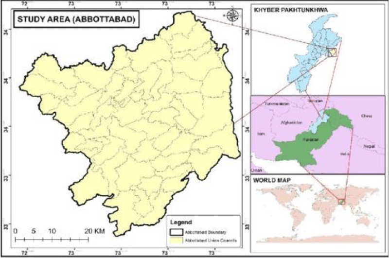
Image 1: Map of study area.
Collections of rust-affected plants were carried out and then infected plants were preserved and submitted to the herbarium of the Department of Botany Hazara University, Mansehra, Pakistan, and then the preserved rust spores were mounted in glycerin jelly and fixed as semi-permanent slides. The spores were measured and photographed under a Leitz HM-LU compound light microscope. At least 30 spores were measured for each spore stage, including the smallest and the largest spores found. Host plants were identified by comparing new collections with the flora of Pakistan and botanical specimens held in the herb. An alcoholic solution of Mercuric Chloride ((Hg Cl2) was used to poison these specimens and it prevented fungal attack and insect damage. With the help of an expert the host of the plants was identified [8].
Survey and collection
The study was conducted on rust fungi in different seasons in 2018. From Thandhiani, District Abbottabad, from selected sites, the plants were collected which were under attack by rust fungi. It was brought under the detailed and closed study according to the instructions of the supervisor. The plants that were healthy and green with flowers or inflorescence and fruits were given special attention during the identification of the exact host. During collection three pictures of every infected plant were taken. Samples were carefully and artistically pressed and then these collected samples were kept among the blotted papers and newspaper. With the passage of time, every day the specimens were exposed to air under strict surveillance, and the newspapers and blotted papers were changed to make it dry. Under the guidance of the supervisor, the above-mentioned process was repeated again and again to make these plants completely dry. For taking clear and high-megapixel pictures, the modern digital camera was used and pictures were taken. An alcoholic solution of Meruric Chloride ((Hg Cl2) was used to poison these specimens and it prevented the fungal attack and insect damage [8]. With the help of an expert the host of the plants was identified. According to the flora of Pakistan, botanical names, family names, and authors' name was corrected. To the herbarium of the Botany department, Hazara University, Mansehra, further study was carried out on these infected plants [9].
Lab work
The Sori part or infected section of the plants was brought under a stereo microscope for study which had a magnification power of 25 to 50. We took at least three pictures with the help of a high megapixel digital camera which was attached to the microscope. All the compulsory equipment and apparatuses were ready in the next step: spirit, spirit lamp, slide, nail lacquer, lectophenole, cover slip, syringe, needle, and tissue paper. In the coming step, we were ready and prepared the slide. We used a needle to take spores for preparing the slide (in the rust fungi case) and mounted it in the lacto phenol. For the semi-permanent slides, we prepared cementing coverslips with the help of nail lacquer, and slight heat due to heating the spore was dispersed. In the coming step, the prepared slide was transferred to the biological microscope and we took from it at least 20 pictures with the help of a digital camera which was attached to the microscope (Nikon YS 100). Lucida camera (Ernst Leitz Wetzlar Germany) was taken and used for the exact drawing of various types of spores and Paraphyses. The spore dimensions were taken by an ocular micrometer (Zeiss Eye Piece Screw Micrometer). For the recording dimension of the spore, extra care was given and it was checked at least twenty pictures of the smallest and biggest spore were taken. After this all the significant data was written in copy and the specimen was described one by one theoretically and then its description was contrasted to the accessible literature for the correct recognition of rust fungi. Then all the infected species were taken and the study was deposited in the Herbarium of the Department of Botany, Hazara University Mansehra, Pakistan [10].
Melampsora euphorbiae (Ficinius and C. Schuib.) (Figure 1, A-H)
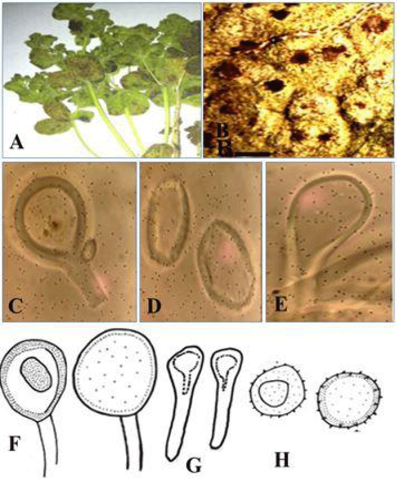
Figure 1: (A) Melampsora euphorbiae Infected host plant. (B) Cross section of an infected leaf showing telium containing teliospores. (D) Uredinospore. (E) Paraphyses. Line drawing of teliospores. Scale bar for A = 2.6 cm, B = 29 mm, C = 14 µm, D = 13.5 µm, E = 7.6 µm, F = 5.6 µm and G = 6.5 µm.
In the aeciospores as well as Spermogonia are totally absent or unknown. Uredia, amphigenous, spread or circinate, yellowish, Uredinospore globosely, sub globosely or ellipsoid, 17. 5 - 20.5 x 13 - 18 µm. Paraphyses capitate, thick-walled 14.3 - 30 µm. Telia sub-epidermal, amphigenous, brownish. Teliospores are cylindrical oblong, yellowish brown, 36 - 66 x 9 – 14 µm.
Material examined: On Euphrbia helioscopia, with stages II and III, Pakistan, Khyber Pakhtunkhwa province, district Abbottabad, Thandiani region, at 2790 m. a. s. l., 15 september, 2018.
Comments: This rust fungus has previously been reported on Euphorbia hypericifolia L. and E. helioscopia L., from Gilgit, Karachi, and Muzaffarabad [11]. It is a new record for the Thandiani region.
Phragmidium barclayi Dietel. (Figure 2, A-F)
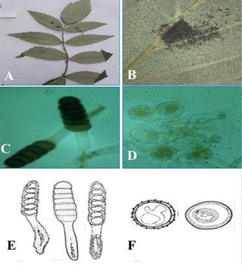
Figure 2: Phragmidium barclayi A. Rubus niveus, B. Telialsori pitcher C. Teliospores D. Uredinospore, A = 1.6 cm, B = 1.3 mm, C = 32.5 µm D = 9 µm, E = 30.5 µm and F = 8.8 µm.
In this case, Barclay shows the uredinal stage as well as Teliospores. But aceia and Spermogonia are absent. Uredinia, light, brown, spread, biaxial, 2 – 3.9 mm. and uredinal spore are mostly ellipsoid and their color is yellow 9.3 – 14. 12 × 13 – 21 µm. germ pores obscured, wall thickness 1.24 – 1.44 µm. telia brown dark, hypophyllous, and scattered about 4mm in diameter, and spores are slightly cylindrical at the base and composed of 3 to 7 cells and the apex is 4.4 – 5.7 µm. In the case of Teliospores about 25 – 34 × 73 – 117 µm, and wall length 2.3 – 3.2 µm. Pedicels are hyaline, 16.6 - 19.3 µm, broad, up to 137 µm long.
Material examined: Pakistan, Khyber Pakhtunkhwa, Abbottabad District, at 2770 m a.s.l., On Rubus niveus II and III stages, 30 September, 2018.
Comment: Phragmidium barclayi has been reported on Rubus lasiocarpus in KP and ChanglaGali by Ahmad [10], Phragmidium barclayi on Rubus spediunculosus is again described from swat. It is a new record for Abbottabad district.
Puccinia ambeguai (Alb.) (Figure 3, A-D)
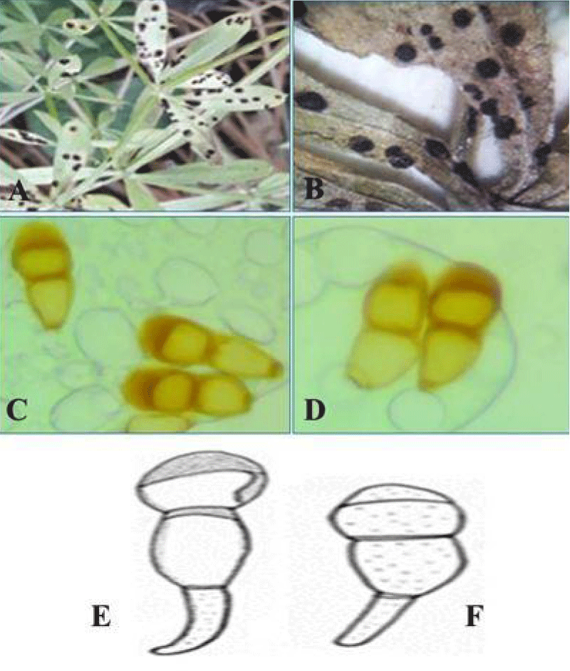
Figure 3: Pucciniia ambegua A Gallium aprine L., B. Telia C. and D. Teliospores scale bar: A = 1.3 cm, B = 1.6 mm, C = 22.1 µm, D = 25.11 µm and Lining drawings of Teliospores, scale bar: A = 25.1 µm.
Spermogonia and aecia, not found. Uredinia hypophyllous, large may be dense, darkly, brownly 1.3 to 1.5 mm. Telia clavate ellipsoidal, apexil, conical, thickened 1.3 to 1.5 mm, teliospores darkly brawny hyaline to pale yellowish, 38. 24 - 57.77 µm × 13. 35 - 20.66 µm. Pedicel 2.35 - 6.35 × 7.88 - 27.97 µm, and hyline.
Material examined: Pakistan, Khyber Pakhtunkhwa, Pakistan, Khyber Pakhtunkhwa, Abbottabad, at 2740 m a.s.l., On Gallium aprine L., II and III stages, October 2018.
Comment: In Gallium aprine plants first of all only the uredinal stage was discovered but here telio stage was also discovered previously it is reported from Murree Ghoregalii by [12] new records for Abbottabad.
Pucciniia calcitraipae DC. (Figure 4, A-F)
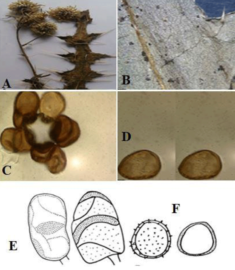
Figure 4: Cirsium vulgare. A. Host Plant. B. Infected leaf showing telium. C. Urediniospores D. Telio-spores scale bar A = 3 cm, B = 1 mm, C = 7 µm, D = 4 µm, A - B Line drawing of A Teliospores B. Uredinia spores scale bars A. = 4 µm B = 2 µm.
In this case, aecia and spermogonia are totally absent. Hypophyllous, Uredinia spread curved, brawny to palely radish. Uredinia - spores globose to ellipsoids. 19 -24 x 23.22 – 27 µm. Telia hypophyllous infrequently, spotted; into groups of various sizes and dark brown color. Teliospores are generally ellipsoidal or fuse-form 15 – 27 x 18 – 40 µm. High and inferior end round, rare case at the septum, also present. Yellow to brown, germ pore apically are sub-apical cells. Pedicle hyaline.
Material examined: Puccinia calcitraipae on Cirsium vulgar with II and III, stages, Pakistan, Khybar Pakhtankhwa Province, District, Abbottabad, Thandiani region, 2600 m. a . s. l., 10 septembar, 2018.
Comment: P. calcitraipae before report on Centaurea bruguieriana (DC.) From Peshawar; on leaves of Cardus edelbergii Rech. f., from Batakundi, Shogran (Kaghan valley), and Kalam (Swat), on leaves of Cirsium arggracanthum Dc,c.Walichii Dc and Cnidus from Maindam, Kalam, Changla Gali, and Shogran [13,14].
Pucciniia mentha Pers. (Figure 5, A-D)
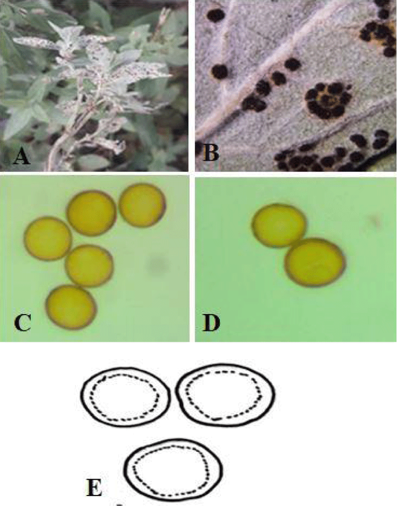
Figure 5: Puccinia menthae A. Mentha longifolia L., B. Telial. C D. Urediniospores. Scale bar: A = 1 cm B = 1 mm C = 10 µm D = 7 µm and E = Lining drawing of Urediniospores scale bar A = 4.2 µm.
Spermogonia and aecia are unknown. Uredinia brown, abaxial, naked, uneven, and spread, Urediniospores hyaline, sub-globose, 18 – 20 × 19 – 28 µm; wall echinulate, 1.4 – 2.3 µm broad, Telia, brownie to black, wall 2- 3 µm thick, verruculose, pedicle little, typically broken.
Material examined: Pakistan, Khyber Pakhtunkhwa, Abbottabad District, Thandiani region, 2730 m a. s. l., On Mentha longifolia, with II, III stage, 8 Sep. 2018.
Comments: P. mentha has been reported on M. Sylvester’s L., from Peshawar, Mingora, Kaghan Valley, Challianwala, Quetta, Poonch, and Muree Hills; on Origanum vulgare L. from ChanglaGali and Kaghan Valley; by [15,16]. Recently it has been reported from Battagram (Fahad 2017). It is the first time reported for the study area.
Puccinia ustalis Bark. (Figures 6, A-D)
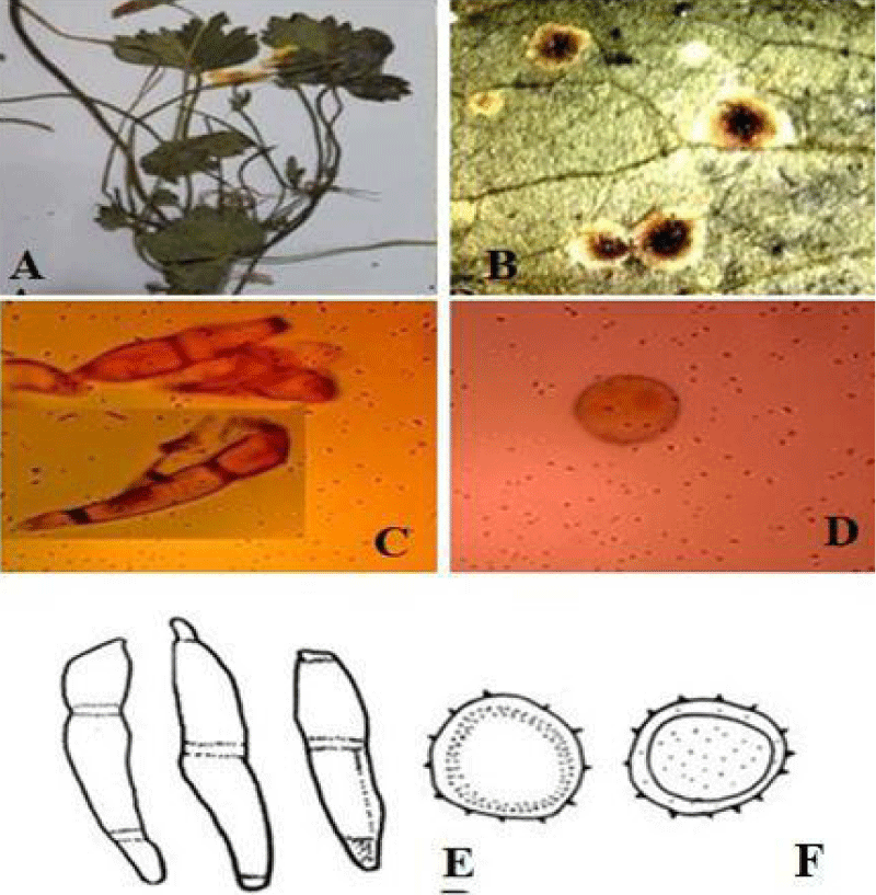
Figure 6: Puccinia ustalis (A). Infected Leaves of Ranunculus muricatus (B). Section of the sorus showing teliospores (C) Uredinospore (D). Teliospores. Scale bar for A = 1.9 cm, B: 5 mm C =15 µm D = 6.5 µm, E, Lining drawings of Teliospores, Uredinospore. scale bar E = 14 µm, F = 7 µm.
Spermogonia, aecia unknown. Uredinia hypophyllous, dark brown to blackish spots, scattered or aggregated in large groups, compact. Urediniospores are ovoid, 9.8 – 23 × 11 - 22 µm, wall thickness 0.6 - 1.7 µm. Telia is usually hypophyllous as dark brown to blackish spots, scattered or aggregated in large groups, compact. Teliospores are mostly two-celled, occasionally 1 or 3 celled teliospores co-exist. ellipsoid, oblong to cylindrical or irregular, conical or rounded above and narrowed below, constricted at the septum, yellowish brown, pale brown basally, 2 celled spores 10–17.2 × 31.6–62.2 µm, smooth,1–1.4 µm thick at the side, epically 2–8 µm thick. Germ pore one per cell, apical or subapical in the distal cell, in a basal cell near the septum, or obscure. Pedicel pale yellow to hyaline, deciduous, 6–9 µm long, deciduous.
Material examined: P. ustalis on Ranunculus muricatus with stage II &III, Pakistan, Khyber Pakhtunkhwa Province, District Abbottabad, Thandiani region, at 2600 m.a.s.l., 15 September 2018.
Comments: P. ustalis has been reported on Anemone obtusiloba D. Don. from Changla Gali and Nathia Gali by Ahmad (1956a, b); on R. diffuses DC. from Dunga Gali KP (NWFP) by [12]and from Kaghan Valley by [17]. Ranunculus muricatus is a new addition to the rust flora of Pakistan on the base of the uredinal stage and a new host.
The goal of the current study project is to learn more about the rust fungi found in the Khyber Pakhtunkhwa Thandhiani region, specifically in District Abbottabad. September and October of 2018 saw the visitation of several carefully chosen locations and the collection of specimens needed for the study. Following a thorough morpho-anatomical search of a number of specimens, six genera—representing six rust species—on six different host vegetation species have been identified. With between 3000 and 4000 species, it is one of the largest groups of fungi. It targets all kinds of vegetation. Gymnosperm, bryophytes, Pteridophytes, and angiosperm may all be impacted. Puccinia is unique in that it can have two or three cells, smooth walls, and a well-developed pedicel. Each Puccinia spore has two or more germ pores and a wall with varying pigmentation [9]. The genus Puccinia is the largest in the current examination, with four species; Ph. Barclayi on Rubus niveus is represented by one species; and Melampsora euphorbiae on Euphrbia helioscopia is represented by one species. Based on the morphology of the spores and the disease pattern, in the current research from Abbottabad, one Taxon M. Euphorbiae, was identified. Previous reports of the rust fungus Euphoribiae hypericifolia from Muzaffarabad, Karachi, and Gilgit [10]. From Charsadda on leaves, Nathiagali, Quetta, Peshawar, Muree, Lahore, Kaghan Valley, Swat, Wazirabad, Challianwala [18]. It was determined that this was a new record for the study area. Ahmad has reported finding Phragmidium barclayi from Rubus lasiocarpus in KP and Changla Gali. Again from SWAT, Phragmidium barclayi on Rubus pedunculosus is described with the most recent illustrations. It's a first for the district of Abbottabad. P. ambegua has been documented on Gallium aprine L.; however, Asghar Ali (1955) only reported its uredinal stage in Muree Hills, Ghoregalii. The fungus's telial stage is a recent addition to Pakistan's rust flora. Additionally, it sets a new record for the Thandiani region's Abbottabad District. Prior research on Centaurea bruguieriana (DC.) Bornm was done by Calcitraipae. A recent addition to Pakistan's rust flora at the base of the uredinal stage is Ranunculus muricatus.
The results of our research indicate that District Abbottabad is extremely wealthy and has a wide variety of fungi, particularly rust. Puccinia, with four species, was found to be the largest genera among the reported genera, followed by Uromyces, with four species. The study's findings indicate that District Abbottabad, which is covered in 70% coniferous forest and is situated in a moist temperate climate, has a huge potential for fungal diversity. Future research in this area is necessary to look for extremely valuable species belonging to other groups.
The authors would like to have collection done in the study research area and lab work is Completed in the Mycology and Plants Pathology lab, Faculty of Health and Biological Science, Department of Botany Hazara University Mansehra Pakistan.
- Cummins GB, Hiratsuka Y. Illustrated genera of rust fungi. 3rd ed, American Phytopathological Society. St. Paul, MN. 2003.
- Mahadevakumar S, Szabo LJ, Eilam T, Anikster Y, Janardhana GR. A New Rust Disease on Wild Coffee (Psychotria nervosa) Caused by Puccinia mysuruensis sp. nov. Plant Dis. 2016 Jul;100(7):1371-1378. doi: 10.1094/PDIS-07-15-0789-RE. Epub 2016 Apr 13. PMID: 30686192.
- Ali B, Sohail Y, Mumtaz AS, Berndt R. Phragmidium punjabense, a new species of rust fungus on Rosa brunonii in the outer Himalayan ranges of Murree, Pakistan. Nova Hedwigia. 2017.
- Ali B, Sohail Y, Tasmia B, Mumtaz AS. Biogeography of rust fungi and their hosts in Pakistan. Science International. 2016; 28(5): 4777–4781.
- Singh AS, Palni UT. Diversity and distribution of rust fungi in Central Himalayan Region. Journal of Phytology. 2011; 3(2): 49-59.
- Pei HM, Hunter T, Royle DJ. Host-Pathogen relationship between Salix and Melampsora sheds light on the parentage of some biomass willows. New Phytologist. 1999; 141: 151-160.
- Bollmann J, Scholler M. Life cycle and life strategy features of Puccinia glechomatis (Uredinales). Mycosccience. 2006; 47: 152-158.
- Haq FUH, Ahmad H, Alam M. Species diversity of vascular plants of Nandiar valley western Himalaya, Pakistan. Pak J Bot. 2010; 42: 213-229.
- Ahmad S, Iqbal SH, Khalid AN. Fungi of Pakistan. Sultan Ahmad Mycological Society of Pakistan. Department of Botany, University of Punjab, Lahore, Pakistan. 1997; 1-248.
- Ahmed S, Iqbal SH, Khalid AN. Fungi of Pakistan. Sultan Ahmad Mycological Society of Pakistan. Department of University of Punjab, Lahore, Pakistan, Quaid–e–Azam Campus, Lahore, Pakistan. 1997; 120–121.
- Ali SI, Qaiser M. A phytogeographical analysis of the Phanerogams of Pakistan and Kashmir. Proc Royal Soc Edinburg. 1986; 89: 89-101.
- Fiaz M, Jabeen S, Imran A, Ahmad H, Khalid AN. Macrolepiota distribution extends to the montana temperate forests of Pakistan. Mycotaxon. 2014; 129(1): 197-208.
- Afshan NS, Khalid AN, Iqbal SH, Niazi AR, Sultan A. Puccinia subepidermalis sp. nov and new records of rust fungi from Fairy Meadows, Northern Pakistan. Mycotaxon. 2009; 110: 173–182.
- Ahmad S, Lodhi SA. Some new or unreported fungi from West Pakistan. Sydowia. 1953; 7(1–4): 266-269.
- Ono Y. Uredinales collected in Kaghan Valley, Pakistan. Cryptogamic Flora of Pakistan. 1992; 1(1): 217-240.
- Okane I, Kakishima M, Ono Y. Uredinales collected in Muree hills, Pakistan. Cryptogamic Flora of Pakistan. 1992; 1(1): 185-196.
- Cummins GB, Hiratsuka Y. Illustrated genera of rust fungi. Third ed. American psychological society. PS Press. Paul, MN. 2003; 1-230.
- Ono Y, Kakishima M. Uredinales collected in Swat Valley, Pakistan. Cryptogamic Flora of Pakistan. 1992; 1(1): 197-216.